Česky
Česky


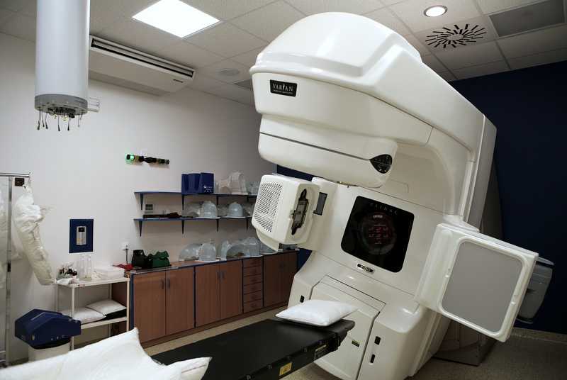
The installation of a new computed tomography (CT) scanner will not only bring better quality images but will also support the possibility of irradiation using the "respiratory gating" technique. Since some lung, liver and subhepatic tumours change their position rhythmically as a result of human breathing, several CT scans in quick succession are necessary to determine their actual location in order to obtain accurate information about their position over time. When treating with conventional techniques, a large safety margin is necessary to ensure the position of the tumour in the irradiated volume throughout the respiratory cycle. With the respiratory gating technique, irradiation only takes place at a certain stage of the respiratory cycle, or the radiation is synchronised with the "moving target volume".This can significantly spare healthy tissue and safely increase the therapeutic radiation dose.
The upgrade will also include the ability to perform so-called image-guided radiotherapy (IGRT). This technique uses imaging during the actual irradiation to provide three-dimensional control of the body position and target volume. Linear accelerators for IGRT are equipped with imaging systems that scan the irradiated area before the actual irradiation. This provides a real-time, up-to-date anatomical view of the irradiated area. By comparing the obtained "image" with the image during treatment planning, it is possible to detect deviations in the settings or changes in the position of the target volume, which are corrected before the actual irradiation starts.This technique allows to refine the irradiation, to increase the dose to the tumour in a targeted way and at the same time to reduce the irradiation of healthy tissues around the tumour. Up to now, this method can only be performed on one device.After the upgrade, it will be possible to perform targeted irradiation on all irradiated patients.
The use of RapidArc technology will also be a significant advance in treatment. This allows highly targeted irradiation of patients in one or two swings of the accelerator arm, reducing the actual irradiation time (in a position that is uncomfortable for some) from 15-25 minutes to approximately 2 minutes.
Last but not least, thanks to the new magnetic resonance it will be possible to perform the so-called MR spectroscopy.This allows not only a detailed anatomical view but also to monitor the concentrations of various substances and to refine the information about the tumour location based on their ratio. This information can be used for targeted dose escalation to the tumour itself, for example in patients with prostate cancer, where the whole prostate is currently irradiated without regard to the actual location of the tumour.
All of these improvements in patient care, not only in the Pardubice Region, which will result in shorter treatment times, increased safety and increased probability of recovery, will be made possible by the implementation of the project "Modernisation and renewal of the equipment of the Radiology Centre Multiscan s.r.o." supported by the European Union from the European Regional Development Fund and implemented under the Integrated Operational Programme through the Ministry of Health. It is also necessary to thank both the Pardubice Regional Hospital and the Pardubice Region for their helpful cooperation in this extensive renewal of necessary technologies.
A chance for your development
Co-financed by the European Union from the European Regional Development Fund